Quick Answer
A dentist distinguishes enamel wear from tooth decay by combining high-magnification visual inspection, tactile probing, and digital X-rays. Enamel wear presents as smooth, hard, cupped-out, or notched yellow dentin across multiple teeth, whereas active decay shows up as soft, sticky, demineralized enamel spots with localized radiographic shadows beneath the surface.
- Enamel wear (attrition, erosion, abrasion) is mechanical or chemical thinning of hard tissue without bacterial infection.
- Tooth decay (caries) is an active bacterial infection that softens and destroys tooth structure from within.
- Worn enamel feels hard and smooth to a dental probe, while active decay feels soft, leathery, and sticky.
- Digital X-rays show clear dark demineralization shadows inside decaying teeth that are absent in surface wear.
Start with the full situation, not one isolated detail
When cold water or sweet food triggers a twinge in your tooth, it is impossible to know what is happening from symptoms alone. Both conditions expose the underlying porous dentin, allowing temperature and osmotic stimuli to reach the dental pulp.
However, treating enamel wear requires desensitization, bite protection, or dietary acid management, whereas treating tooth decay requires removing infected bacterial tissue and placing a sealed restoration.
The useful question is not only whether sensitivity from enamel wear versus tooth decay is common. It is whether the pattern, timing, location, and associated symptoms point to something that needs prevention, monitoring, repair, or urgent treatment. A dental assessment combines what you have noticed with the condition of the teeth, gums, bite, and existing dental work.
What may explain sensitivity from enamel wear versus tooth decay
Several explanations can produce a similar experience. Appearance or symptoms at home can narrow the conversation, but they cannot reliably confirm the diagnosis. The dentist looks for a finding that explains the whole pattern rather than choosing treatment from one symptom.
Tell the dental team what changed, when it started, whether it is improving or recurring, and what makes it better or worse. That history helps the examination focus on the most relevant teeth and tissues while still checking for another source.
| Possible factor | Why it may matter |
|---|---|
| Acid erosion and toothbrush abrasion | Dietary acids, reflux, or aggressive scrub-brushing wearing away enamel collars at the gumline. |
| Active interproximal or occlusal caries | Bacterial acid demineralizing enamel and rotting the underlying dentin layer. |
| Attrition from bruxism clenching | Flattened chewing surfaces where enamel has been worn away by tooth-on-tooth friction. |
| Arrested non-active decay vs active lesion | An old dark stain that has remineralized and hardened versus an active softening cavity. |
What the dentist may check
The appointment begins with your symptoms, dental history, medicines, allergies, health conditions, and previous treatment. The clinical examination then tests the possibilities that fit sensitivity from enamel wear versus tooth decay. An X-ray or another diagnostic step may be used only where it adds useful information.
No single test answers every dental question. Visual findings, gum measurements, bite response, sensitivity testing, imaging, and comparison with nearby teeth are interpreted together. If the source is uncertain, the safest plan may include review or referral before irreversible treatment.
| Check | What it helps establish |
|---|---|
| Tactile hardness probing with fine explorer | Testing whether the sensitive surface is hard and glassy (wear) or soft and sticky (active decay). |
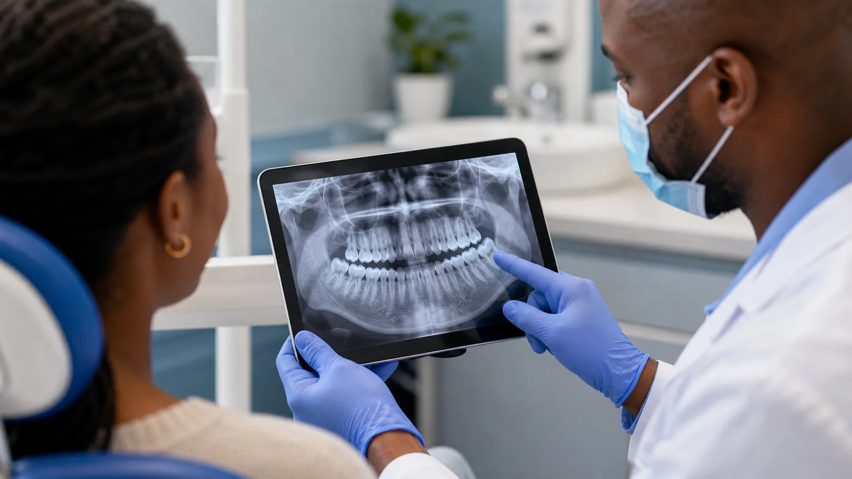
| Digital bitewing and periapical X-rays | Checking for internal radiolucency (dark shadow) indicating bacterial penetration into dentin. |
| Caries detection dye or laser fluorescence | Using diagnostic aids to highlight active bacterial collagen breakdown in questionable fissures. |
| Dietary and lifestyle risk assessment | Reviewing soda, citrus, reflux, brushing pressure, and night-time clenching habits. |
How the next step is chosen
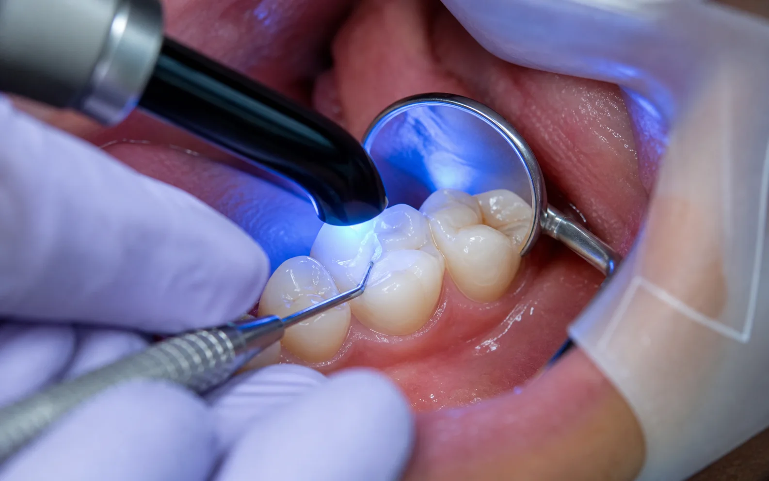
If the area is hard and bacterial infection is ruled out by X-rays, non-invasive desensitizing and habit adjustments protect the tooth. If bacterial decay is active, prompt filling stops the cavity from reaching the nerve.
The recommended option should match the diagnosis, the amount of healthy tooth and gum support, the urgency, your wider health, and whether the result can be maintained. Ask the dentist to distinguish treatment needed to control disease from elective improvements that can safely wait.
A complete plan should include any temporary stage, definitive procedure, final restoration, and review. If care is staged, confirm what protects the area between visits and which change means the next appointment should be brought forward.
| Possible next step | When it may enter the discussion |
|---|---|
| For Enamel Wear: Desensitizing varnish & sealants | Sealing exposed tubules, modifying brushing technique, and providing custom night guards. |
| For Enamel Wear: Cervical bonding | Placing tooth-colored composite or glass ionomer to fill deep mechanical notches and restore enamel thickness. |
| For Active Decay: Minimally invasive composite filling | Excavating infected bacterial tissue and placing a biomimetic bonded filling. |
| For Advanced Decay: Pulp therapy or crown | Required if decay has penetrated near or into the dental pulp nerve chamber. |
Turn the diagnosis into a complete care plan
A useful plan for sensitivity from enamel wear versus tooth decay should connect every recommended step to a finding from the examination. Ask the dentist to separate what controls active disease or immediate risk from what restores function, improves appearance, or prevents recurrence. That distinction makes it easier to understand why one step comes before another.
Confirm whether treatment can be completed in one appointment or requires a temporary stage, laboratory work, healing, specialist input, or a final restoration. If several visits are involved, ask what protects the tooth or gum between them, which activities or foods should be limited, and whom to contact if the temporary stage changes.
Long-term value also depends on maintenance. Discuss how the area will be cleaned, which review findings show that treatment is working, what can fail over time, and whether future repair is possible. Where cost affects timing, request a written sequence that identifies the urgent phase, the definitive phase, and any later elective or replacement decision.
Record the diagnosis and agreed sequence in language you understand. If more than one option is suitable, compare expected durability, maintenance, recovery, limitations, and the likely result of delaying each choice. A clear written plan also helps another clinician understand what has already been assessed if you need urgent care or continue treatment at a different practice.
What to do before the appointment
Switch to a soft toothbrush and use non-abrasive sensitivity toothpaste. If consuming acidic foods or drinks, rinse with water and wait 30 minutes before brushing.
Home care should protect the area without hiding a worsening problem. Avoid scraping, forcing tools, applying chemicals to teeth or gums, or using someone else’s prescription. Use only medicines that are safe for you and follow the label or advice from a qualified healthcare professional.
Keep the appointment even if the symptom settles. A changing symptom is still useful evidence, and temporary improvement does not prove that decay, inflammation, infection, a crack, or a gum problem has resolved.
- Note whether the sensitive area feels rough, notched, or hollow to your tongue.
- Notice if the sensitivity is worse with cold, hot, or sweet food.
- Consider if you have a history of acid reflux, lemon water consumption, or hard brushing.
- Avoid scraping the sensitive area with toothpicks or metal tools at home.
Questions that make the plan clearer
Good questions connect the proposed care to a specific finding. Ask what the dentist can confirm today, what remains uncertain, and what would change the recommendation. This is more useful than comparing procedure names without knowing whether they address the same diagnosis.
Also ask about the full sequence, likely review point, maintenance, alternatives that are clinically suitable, and the consequences of waiting. Where cost matters, request an estimate that covers the complete plan rather than only the first urgent or temporary stage.
- Is the sensitive area caused by active bacterial decay or by mechanical enamel wear?
- Do my digital X-rays show any decay between the teeth that cannot be seen with the eye?
- Can this be treated with fluoride and desensitizing glazes, or is a filling necessary?
- What steps should I take to prevent further enamel erosion across my other teeth?
When to arrange faster care
Move the appointment forward when symptoms are worsening, affecting sleep or eating, or accompanied by swelling, pus, a bad taste, fever, trauma, uncontrolled bleeding, or increasing difficulty opening the mouth. Describe these changes when booking so that the practice can triage the situation.
Difficulty breathing, swallowing, or speaking, extensive mouth or neck swelling, swelling affecting an eye, severe facial trauma, confusion, or uncontrolled bleeding can require emergency medical help. Do not wait for an ordinary dental appointment when airway, vision, consciousness, or general health may be affected.
Without emergency signs, persistent or recurring sensitivity from enamel wear versus tooth decay still deserves assessment. Early diagnosis usually gives the dentist more information and may preserve more conservative choices than waiting for a crisis.
- Pain shifts from cold-triggered twinges to spontaneous throbbing without any stimulus.
- A visible brown, grey, or black hole develops in the tooth.
- Food constantly packs tightly into an open hollow on the tooth.
Internal Links
Related dental care
Sources
Useful information
Written by
Dr. Kholofelo Machaba-SelatoleChief Dentist & Practice Director
Dr. Kholofelo Machaba-Selatole leads Smile On Dental & Aesthetic Studio with a warm, patient-focused approach to family, restorative, cosmetic, and orthodontic care.
View Profile