
Quick Answer
Yes, a painless dark spot can still be tooth decay because early cavities often cause no symptoms. It can also be external stain, a naturally dark groove, or discolouration around an existing filling. Do not pick at the spot or cover it with whitening products. A dentist can inspect the surface, check whether it is soft or structurally changed, review nearby fillings and gums, and use an X-ray when the location cannot be fully assessed. Book sooner if the spot grows, traps food, becomes sensitive, or sits beside a cracked or broken area.
- Pain is a late or unreliable way to decide whether decay is present.
- Colour alone cannot distinguish stain, a natural groove, decay, or restoration changes.
- Early mineral loss may be managed differently from a cavity with permanent structural damage.
- Cleaning, whitening, and filling treatment have different purposes.
Why decay can be painless
Tooth decay develops over time as repeated acid exposure removes minerals from enamel. Early changes can occur without pain because the outer enamel does not respond like the inner tissues of the tooth. A cavity may remain quiet until it becomes larger or reaches a more sensitive layer.
The absence of pain is therefore reassuring only in a limited sense. It does not show whether the surface is healthy. A small problem found during a routine examination may allow more conservative treatment than waiting for sensitivity or toothache.
A dark mark is not automatically decay either. Staining can collect in grooves, and natural anatomy can create shadows. Diagnosis depends on the surface, location, progression, surrounding tooth structure, and sometimes imaging.
What else can look like a dark spot
External stain from foods, drinks, or tobacco can mark enamel and collect in pits and grooves. Tartar can also become stained. An older filling may have a dark edge, or the tooth may show a line or shadow that is not an active cavity.
A crack, chipped area, or food trap changes the concern because it may expose weaker tooth structure or allow plaque to remain. A spot between teeth is difficult to assess in a mirror and may only be visible clinically or on an appropriate X-ray.
Whitening products cannot diagnose or repair these causes. They do not remove tartar, seal decay, or fix a filling margin. A cleaning may remove surface deposits, but a structural problem needs a restorative assessment.
| Possible explanation | What distinguishes it |
|---|---|
| Surface stain | May sit on intact enamel and respond to professional cleaning. |
| Stained groove | Needs examination to decide whether the groove is sound or decayed. |
| Early mineral loss | May need prevention and monitoring when the surface is still intact. |
| Cavity | Permanent structural damage may require a filling or another restoration. |
| Old filling edge | The dentist checks seal, tooth structure, symptoms, and imaging where needed. |
How the dentist checks the spot
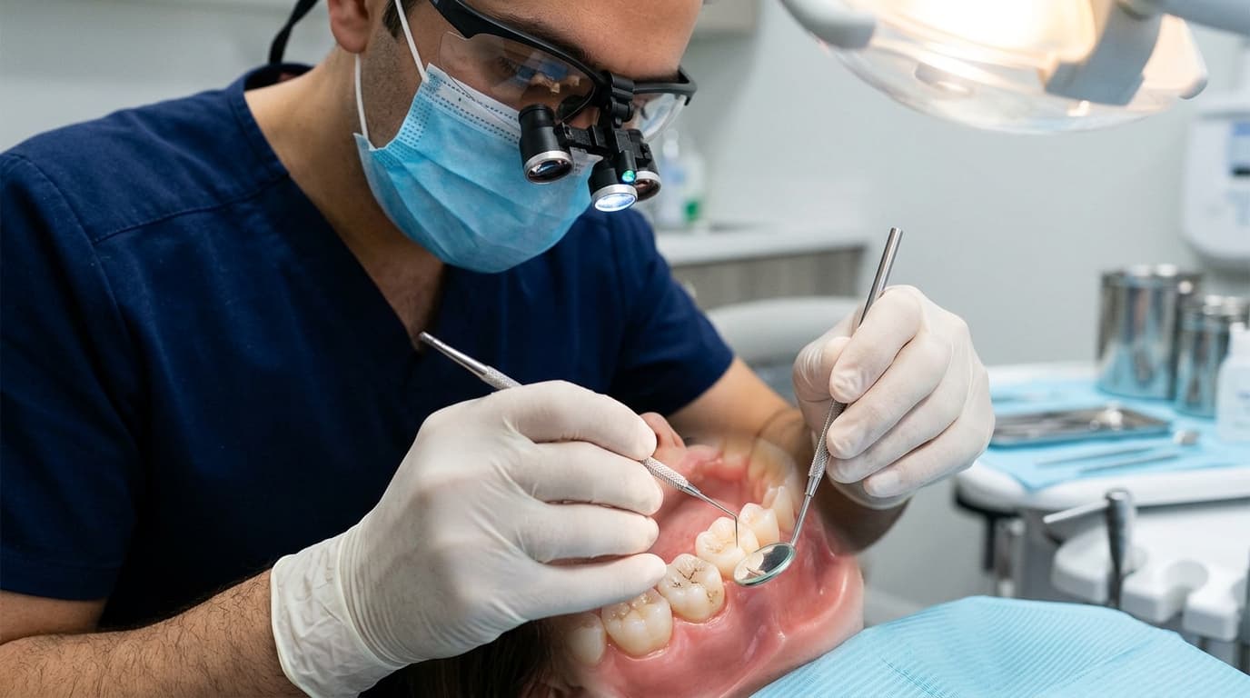
The dentist examines the tooth under good lighting, dries the surface, checks nearby gums and restorations, and considers the shape and location. The goal is to determine whether the enamel is intact, whether food or plaque is being trapped, and whether the area has changed.
An X-ray may be recommended when decay could be between teeth, below a filling, or deeper than the visible surface suggests. Not every surface mark needs an image, and not every early lesion is clearly represented on an X-ray. Findings are combined rather than relying on one test.
Tell the dentist when you first noticed the spot and whether it has grown, become rough, or developed sensitivity. A photograph taken under similar lighting can help you track change, but home photographs do not replace clinical diagnosis.

Early decay and a cavity are not managed the same way
When enamel has lost minerals but the surface is still intact, preventive measures may help stop or reverse the early process in selected cases. Fluoride, better plaque removal, and changes to the frequency of sugary or starchy foods and drinks can support the plan.
Once tooth structure has broken down into a cavity, the damage is permanent and the dentist generally needs to repair it. The size, depth, tooth position, remaining strength, and symptoms influence whether a filling or a stronger restoration is suitable.
Do not use the possibility of prevention as a reason to self-diagnose. The important distinction is whether the surface is intact and whether the lesion is active. That judgement requires a dental examination.
- Confirm whether the mark is stain, early decay, a cavity, or a restoration issue.
- Assess cavity risk and the areas where plaque remains.
- Use prevention and monitoring when clinically appropriate.
- Restore permanent structural damage before it becomes larger.
- Review the tooth and daily routine over time.
What you can change before the appointment
Brush twice daily with fluoride toothpaste and clean between teeth with floss or interdental brushes. Do not scrape the mark with sharp tools, scrub aggressively, or apply bleach products.
Reduce the frequency of sugary and starchy snacks and sweet drinks. Grouping exposures with meals gives saliva and fluoride a better opportunity to protect enamel.
If the tooth becomes sensitive, write down what triggers it and how long the feeling lasts. That information helps the dentist evaluate whether the deeper layers of the tooth are involved.
If the dentist recommends monitoring, define the review point
Monitoring should be an active plan rather than an indefinite delay. Ask what the dentist believes the spot represents, which features make observation appropriate, and when the area should be checked again. The record may include its location, surface condition, photograph, or relevant imaging.
Your part is to maintain fluoride exposure, control plaque, and follow the recommended pattern for snacks and drinks. Tell the dentist if the mark changes before the planned review. A new rough edge, food trap, sensitivity, floss tearing, or visible growth can alter the decision.
At review, the dentist compares the current surface with the earlier findings. Stability may support continued prevention and monitoring; progression or structural breakdown may support restorative treatment. This defined checkpoint helps avoid both unnecessary early treatment and avoidable delay.
If you see another dentist before that date, describe the monitoring plan and share previous images where available. Consistent records make change easier to judge.
When to move the appointment forward
Book sooner if the mark is growing, the surface feels broken, food catches, floss tears, or the tooth becomes sensitive to cold, sweets, or biting. A lost or cracked filling also deserves assessment before more tooth structure breaks.
Pain, swelling, pus, a bad taste, or severe night-time symptoms can indicate that the problem is no longer a routine painless spot. Tell the practice about those symptoms so the urgency can be assessed.
The most useful approach is early identification. If the spot is only stain, you gain reassurance and appropriate cleaning advice. If it is decay, the dentist can plan care before pain becomes the first signal.
Internal Links
Related dental care
Sources
Useful information

Written by
Dr. Kholofelo Machaba-SelatoleChief Dentist & Practice Director
Dr. Kholofelo Machaba-Selatole leads Smile On Dental & Aesthetic Studio with a warm, patient-focused approach to family, restorative, cosmetic, and orthodontic care.
View Profile