Quick Answer
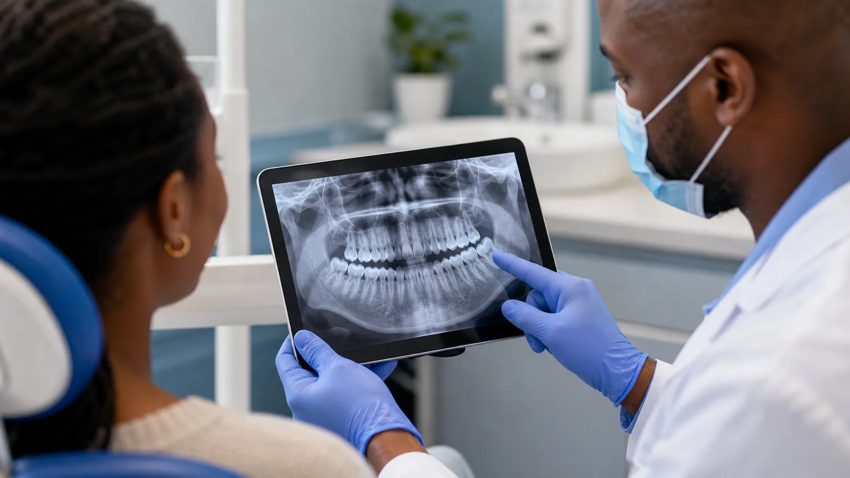
The dentist may recommend an X-ray because the visible part of a problem does not show its full depth or effect on roots and supporting bone. Imaging can help assess decay between teeth or beneath a restoration, infection near a root, bone levels, tooth position, or a fracture-related concern. It should be prescribed only when the expected diagnostic information can affect care. Ask which question the image is intended to answer and how the result could change the plan.
- Clinical examination and X-rays provide different information.
- A visible problem can extend into areas that cannot be inspected directly.
- Images should be selected for a diagnostic reason, not taken automatically.
- A normal image does not exclude every crack or soft-tissue problem.
Start with the full situation, not one isolated detail
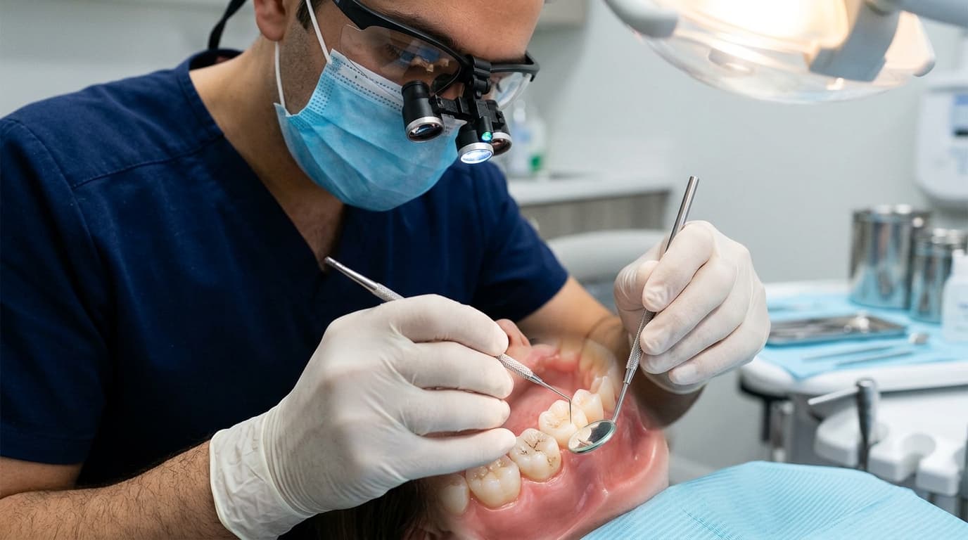
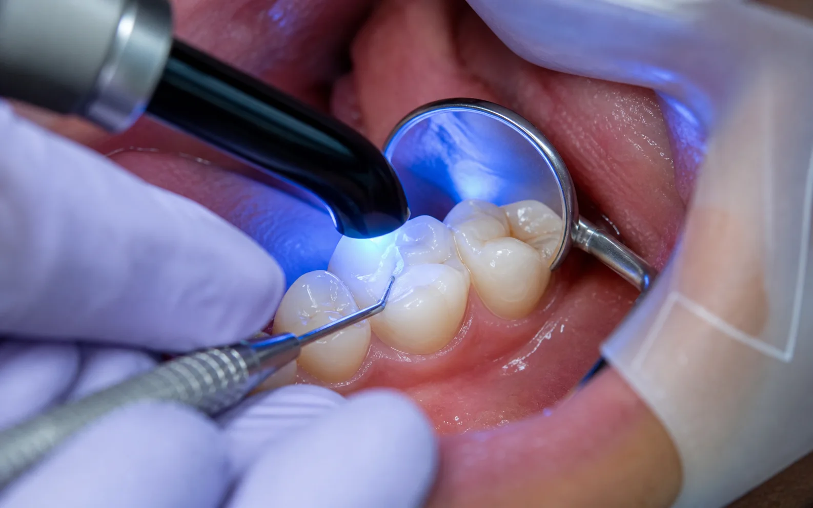
A dentist can see enamel, exposed restoration edges, gums, and part of a fracture, but roots and most supporting bone are covered by tissue. Decay between teeth or beneath a crown may also extend beyond the visible mark.
The image does not replace the examination. It adds a different view that can help judge restorability, infection, tooth position, bone support, and whether a proposed procedure is proportionate.
The useful question is not only whether using an X-ray for a problem that is already visible is common. It is whether the pattern, timing, location, and associated symptoms point to something that needs prevention, monitoring, repair, or urgent treatment. A dental assessment combines what you have noticed with the condition of the teeth, gums, bite, and existing dental work.
What may explain using an X-ray for a problem that is already visible
Several explanations can produce a similar experience. Appearance or symptoms at home can narrow the conversation, but they cannot reliably confirm the diagnosis. The dentist looks for a finding that explains the whole pattern rather than choosing treatment from one symptom.
Tell the dental team what changed, when it started, whether it is improving or recurring, and what makes it better or worse. That history helps the examination focus on the most relevant teeth and tissues while still checking for another source.
| Possible factor | Why it may matter |
|---|---|
| Hidden depth of decay | Shows whether a visible lesion may extend farther into the tooth. |
| Root or bone involvement | Assesses structures that cannot be seen directly. |
| Disease under dental work | May reveal changes beneath a filling or crown. |
| Position and anatomy | Supports planning around roots, nearby teeth, and surrounding structures. |
What the dentist may check
The appointment begins with your symptoms, dental history, medicines, allergies, health conditions, and previous treatment. The clinical examination then tests the possibilities that fit using an X-ray for a problem that is already visible. An X-ray or another diagnostic step may be used only where it adds useful information.
No single test answers every dental question. Visual findings, gum measurements, bite response, sensitivity testing, imaging, and comparison with nearby teeth are interpreted together. If the source is uncertain, the safest plan may include review or referral before irreversible treatment.
| Check | What it helps establish |
|---|---|
| Clinical finding first | Defines the problem and the reason imaging may help. |
| Previous images | May provide adequate recent information or useful comparison. |
| Image type and area | Targets the smallest useful view for the diagnostic question. |
| Result with examination | Combines imaging with symptoms and clinical tests. |
How the next step is chosen
The decision is based on expected benefit. The dentist should consider symptoms, age, disease risk, previous images, the proposed treatment, and whether the information can influence diagnosis or planning. More images are not automatically better.
The recommended option should match the diagnosis, the amount of healthy tooth and gum support, the urgency, your wider health, and whether the result can be maintained. Ask the dentist to distinguish treatment needed to control disease from elective improvements that can safely wait.
A complete plan should include any temporary stage, definitive procedure, final restoration, and review. If care is staged, confirm what protects the area between visits and which change means the next appointment should be brought forward.
| Possible next step | When it may enter the discussion |
|---|---|
| Proceed without an image | May be suitable when imaging would not change care. |
| Take a targeted dental X-ray | Answers a local question about the tooth or supporting tissue. |
| Use a wider view | May be appropriate for position, development, or multiple structures. |
| Refer for specialised imaging | Reserved for selected questions that need more detail. |
Turn the diagnosis into a complete care plan
A useful plan for using an X-ray for a problem that is already visible should connect every recommended step to a finding from the examination. Ask the dentist to separate what controls active disease or immediate risk from what restores function, improves appearance, or prevents recurrence. That distinction makes it easier to understand why one step comes before another.
Confirm whether treatment can be completed in one appointment or requires a temporary stage, laboratory work, healing, specialist input, or a final restoration. If several visits are involved, ask what protects the tooth or gum between them, which activities or foods should be limited, and whom to contact if the temporary stage changes.
Long-term value also depends on maintenance. Discuss how the area will be cleaned, which review findings show that treatment is working, what can fail over time, and whether future repair is possible. Where cost affects timing, request a written sequence that identifies the urgent phase, the definitive phase, and any later elective or replacement decision.
Record the diagnosis and agreed sequence in language you understand. If more than one option is suitable, compare expected durability, maintenance, recovery, limitations, and the likely result of delaying each choice. A clear written plan also helps another clinician understand what has already been assessed if you need urgent care or continue treatment at a different practice.
What to do before the appointment
Bring previous images if another practice has taken them recently and tell the dentist if you are or may be pregnant. Do not decline needed diagnosis only because the surface problem looks obvious; instead, ask for a clear explanation of the purpose and alternatives.
Home care should protect the area without hiding a worsening problem. Avoid scraping, forcing tools, applying chemicals to teeth or gums, or using someone else’s prescription. Use only medicines that are safe for you and follow the label or advice from a qualified healthcare professional.
Keep the appointment even if the symptom settles. A changing symptom is still useful evidence, and temporary improvement does not prove that decay, inflammation, infection, a crack, or a gum problem has resolved.
- Bring available recent dental records and images.
- Share pregnancy and complete health information.
- Ask what structure cannot be assessed visually.
- Ask how the image could change the treatment plan.
- Confirm whether another recent image is adequate.
Questions that make the plan clearer
Good questions connect the proposed care to a specific finding. Ask what the dentist can confirm today, what remains uncertain, and what would change the recommendation. This is more useful than comparing procedure names without knowing whether they address the same diagnosis.
Also ask about the full sequence, likely review point, maintenance, alternatives that are clinically suitable, and the consequences of waiting. Where cost matters, request an estimate that covers the complete plan rather than only the first urgent or temporary stage.
- What exact question will this X-ray answer?
- Which type of image is being recommended and why?
- Do you have a recent image for comparison?
- Could the result change whether the tooth is restorable?
- What are the limits of the image?
When to arrange faster care
Move the appointment forward when symptoms are worsening, affecting sleep or eating, or accompanied by swelling, pus, a bad taste, fever, trauma, uncontrolled bleeding, or increasing difficulty opening the mouth. Describe these changes when booking so that the practice can triage the situation.
Difficulty breathing, swallowing, or speaking, extensive mouth or neck swelling, swelling affecting an eye, severe facial trauma, confusion, or uncontrolled bleeding can require emergency medical help. Do not wait for an ordinary dental appointment when airway, vision, consciousness, or general health may be affected.
Without emergency signs, persistent or recurring using an X-ray for a problem that is already visible still deserves assessment. Early diagnosis usually gives the dentist more information and may preserve more conservative choices than waiting for a crisis.
- Facial swelling, fever, pus, or severe worsening pain
- Recent trauma with movement or displacement
- A rapidly breaking tooth or restoration
- Difficulty breathing, swallowing, or opening the mouth
Internal Links
Related dental care
Sources
Useful information

Written by
Dr. Kholofelo Machaba-SelatoleChief Dentist & Practice Director
Dr. Kholofelo Machaba-Selatole leads Smile On Dental & Aesthetic Studio with a warm, patient-focused approach to family, restorative, cosmetic, and orthodontic care.
View Profile